Platelets
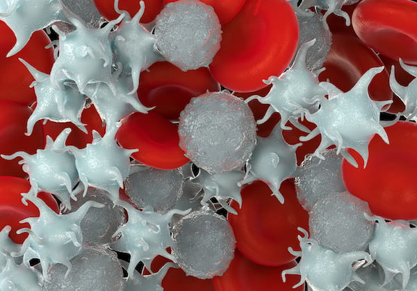
Platelets are small discoid cells of 1-3 µm in diameter that circulate in the blood at a concentration of 1.4-4 x 10⁸/ml. The most well-known function of platelets is in hemostasis, where they are responsible for the formation of a vascular plug to prevent excessive blood loss following injury. More recently, a role for platelets in host inflammatory and immune responses has emerged.
Platelet formation and structure
Platelets are formed in the bone marrow from megakaryocytes, which are one of the eight main blood cell types produced from hematopoietic stem cells. Mature megakaryocytes form structures known as proplatelets. Cellular contents, apart from the nucleus, are distributed into these fragments, resulting in the production of several thousand platelets from each megakaryocyte. They do not have a nucleus, differentiating them from most circulating cells, but the youngest ‘reticulated’ platelets still contain a small amount of RNA, and can consequently be identified using thiazol orange staining and subsequent flow cytometric analysis.1 As this immature state has a short lifespan of only one day, the proportion of reticulated platelets can serve as an indicator for megakaryopoiesis in the bone marrow. Mature platelets circulate in the blood for approximately nine days, before being removed by the spleen.
Each platelet’s plasma membrane contains a high number of diverse glycoprotein receptors and signaling proteins, which allow them to respond rapidly to stimulation. Below this plasma membrane is the microtubule ring that gives resting platelets their unique disc shape. The platelet cytoplasm contains several types of secretory granules, which are released when they are activated, and contain a range of factors that further support thrombus formation and wound repair, such as fibrinogen, von Willebrand factor (vWF), fibronectin, ADP, 5-HT, chemokines, and growth factors.
Platelets in hemostasis and thrombosis
Platelets circulate in the blood in a resting state but undergo rapid activation in response to damage to the vascular wall. When damaged, endothelial cells release vWF, and sub-endothelial cell proteins – primarily collagen – are exposed at the site of injury. Collagen and vWF bind to receptors on the platelet surface, triggering a variety of responses including adhesion, shape change, spread, enhanced procoagulant activity, the release of vascular contents, and aggregation. This results in the formation of a primary hemostatic plug, which stops bleeding and helps to reduce infection following injury.
Platelet activation in diseased blood vessels leads to thrombotic events, including myocardial infarction and stroke. Understanding the processes underlying platelet activation is therefore essential in identifying novel antithrombotic drug targets.
Thrombocytopenia is a condition characterized by abnormally low levels of platelets. There are a number of possible causes, including bone marrow disorders, immune system problems or side effects of certain medications. Thrombocytopenia is often mild, with few signs or symptoms but, in some cases, the number of platelets can be low enough to cause dangerous bleeding.
Emerging research – platelets in immunity and inflammation
Platelet activity in thrombosis and hemostasis is generally well characterized, however, recent research has focused on the emerging role of platelets in a number of other physiological processes, including host defense2 and tissue repair,3 which themselves are involved in cancer metastasis4 and inflammatory disorders, such as rheumatoid arthritis.5 Research into the receptors mediating these pathological functions, and their underlying mechanisms, are therefore a major focus of current platelet research. There is also an increasing understanding of platelet-derived extracellular vesicles (PEVs), which are released when platelets are activated and are implicated in inflammation.5 These are the most abundant extracellular vesicles found in human blood, and early indications suggest that they may represent an important differentiating analyte of significant therapeutic interest.
Tools to study platelets
Culture methods
It is not possible to culture platelets as they are not capable of cell division. Platelets are typically collected in blood samples for use in functional assays, and can be used for experiments within whole blood, platelet rich plasma (PRP), or as ‘washed platelets’, where platelets are extracted from plasma by centrifugation and resuspended in buffer. Once isolated, platelets are very sensitive to changes in temperature, pH, and physical shock, which can cause them to aggregate, and therefore careful attention must be paid to their storage.
Cell counting
Platelet counts can be performed with an automated hematology analyzer, or manually with a hemocytometer. Platelets may be counted to monitor or diagnose diseases, or to look for the cause of abnormal bleeding or clotting.
Functional assays
- Platelet aggregation: Platelet aggregation studies can be used to study agonists and antagonists of platelet activation, and their underlying mechanisms. In its simplest format, agonist is added to PRP or washed platelets in a platelet aggregometer, and aggregation is measured by the change in light passing through the sample. To better mimic platelet activation in the body, platelets can be flowed over agonists at shear rates that mimic physiological conditions and the resulting aggregates viewed by microscopy. The adhered and aggregated platelets can subsequently be labelled for fluorescence imaging.
- Flow cytometry: Flow cytometry can provide detailed molecular information on platelet phenotype and function. Platelets can be analyzed in their resting state, or following activation, based on differential surface markers expressed following stimulation. It may be necessary to fix the sample depending on how quickly analysis will be carried out.
Cell markers
Platelet activation can be monitored based on the presence of different surface markers in the resting and activated state. The reduction or absence of certain markers can also be useful for disease diagnosis.
Overview of the specific markers for resting and activated platelets
| Surface Marker | Alternative Names | Location |
|---|---|---|
| Resting Platelets | ||
| CD9 | TSPAN-29, MIC3, BA2, DRAP-27, MRP-1 | Cell surface |
| CD29 | Integrin β1, ITGB1, Fibronectin receptor subunit beta, FNRB, GPIIA, MDF2, MSK12, VLAB | Cell surface |
| CD31 | PECAM-1, Platelet endothelial cell adhesion molecule | Cell surface |
| CD36 | Fatty acid translocase, Glycoprotein IIIb, GP3B, GP4, GPIV, PASIV, SCARB3 | Cell surface |
| CD41 | Integrin αIIb, GPalpha IIb, ITGA2B, GP2B, GTA | Cell surface |
| CD42a | Platelet glycoprotein IX, GPIX, GP9 | Cell surface |
| CD42b | Platelet glycoprotein Ib alpha chain, BSS, GP1B, GPIbα | Cell surface |
| CD61 | Integrin β3, Integrin beta-3, ITGB3, GP3A | Cell surface |
| GPVI | Cell surface | |
| Activated Platelets | ||
| CD62P | P-selectin, SELP, LECAM3, GRMP, PADGEM | α-granules, moves to cell surface on activation |
| CD63 | LAMP-3, Granulophysin, MLA1, ME491 | Dense granules, moves to cell surface on activation |
| CD107a | LAMP-1 | Lysosomes, moves to cell surface on activation |
| CD107b | LAMP-2 | Lysosomes, moves to cell surface on activation |
| CD154 | CD40 ligand, Gp39, TRAP, HIGM1 | Displayed on activated surface and released as soluble form (sCD154) |
| Activated IIb/IIIa | Cell surface. Undergoes conformational change on activation that can be measured by specific antibodies eg. PAC-1 | |
References
1. Hoffmann JJ. (2014) Reticulated platelets: analytical aspects and clinical utility. Clin Chem Lab Med. 52(8):1107-17. https://www.degruyter.com
2. Cloutier N, Allaeys I et al. (2018) Platelets release pathogenic serotonin and return to circulation after immune complex-mediated sequestration. Proc Natl Acad Sci U S A. 115(7):E1550-E1559. https://www.pnas.org
3. Gawaz M and Vogel S (2013) Platelets in tissue repair: control of apoptosis and interactions with regenerative cells. Blood. 122(15):2550-4. https://ashpublications.org
4. Lucotti S and Muschel RJ (2020) Platelets and Metastasis: New Implications of an Old Interplay. Front. Oncol. 10:1350. https://www.frontiersin.org
5. Boilard E, Nigrovic PA et al. (2010) Platelets Amplify Inflammation in Arthritis via Collagen-Dependent Microparticle Production. Science. 327(5965): 580-583. https://www.science.org
